

HOW DOES THE EYE WORK?
Vision is one of the greatest miracles of the body and arguably the most relied upon special sense. Of vision, Leonardo Da Vinci once said, “The eye, which is said to be the window of the soul, is the principal means by which the brain’s sensory receptor may fully and magnificently contemplate the infinite works of nature” (1). As such, it is not surprising that blindness consistently ranks among the most feared conditions in America.
The structure and function of the eye can be compared to an analog camera where the front compartment of the eye, consisting of the cornea and the lens, focuses light just like the lens of a camera. Light is focused onto the retina, in the back of the eye, just as it’s focused onto the film of a camera. After the retina receives this focused light, it transforms the various colors and shades into a language that the brain can interpret as the picture of our lives. The macula is a specialized area near the center of the retina that is responsible for the center of vision. It could be compared to the most sensitive are of camera film and is what allows for high-resolution vision needed for reading, focusing on distant objects, and completing most tasks.
In order for the retina to receive light and transmit related signals to the brain properly, an underlying tissue, known as the retinal pigment epithelium (RPE) supplies nutrients to and helps clear away waste products from other retinal cells. The RPE also creates suction to keep the other layers of the retina in place. The RPE, located under the macula, or the light receptors above it are often the site of initial damage in the development of a condition known as age-related macular degeneration (2). Damage to the RPE or nearby retinal tissue it is thought to trigger inflammation and subsequent formation of new retinal cells. This inflammation and cell build-up are believed to be the primary driver of age-related macular degeneration. Because the RPE is the primary support for the light sensing cells above it, when it dies, the retina can slowly die as well, causing it to lose some of its light sensing capabilities.
RISK FACTORS FOR AGE-RELATED MACULAR DEGENERATION
Macular degeneration is defined as a multifactorial condition. Its development is influenced by genetic (heritable) factors as well as environmental factors. The top 5 risk factors are:
- Age (#1 risk factor)
- Smoking
- European ancestry (lighter colored eyes allow more light into the retina thereby increasing the risk of UV damage)
- Hypertension
- Obesity (3)
SYMPTOMS OF AGE-RELATED MACULAR DEGENERATION
Age-related macular degeneration (ARMD) eventually leads to blurred vision or complete loss of vision in the center of the visual field. This does not result in complete blindness, but instead results in a loss of central vision that makes it hard to recognize faces, drive, read or perform other activities of daily life. There are three stages of age-related macular degeneration.
The first two stages are based upon the degree of cellular debris, or drusen, present in or under the retina. Because the retinal pigment epithelium normally functions to clear the retina of debris, the presence of drusen usually indicates that 1) the RPE is dysfunctional 2) there is significant damage to the retina or 3) there is dysfunction or damage to both the RPE and the retina (4). Regardless of the cause, an inability of the retinal pigment epithelium to clean up either normal or abnormal cellular waste from the retina leads to impairment in the ability of the retina to receive light. People with early-stage ARMD may, however, have no noticeable symptoms.
The third stage is defined by either retinal pigment epithelium scarring or neovascularization. Neovascularization is the process by which new blood vessels form to bring extra nutrients to the dying tissue and to take away the waste debris. Neovascular age-related macular degeneration is often simply termed wet ARMD, while all other forms of the disease are called dry ARMD. New blood vessels associated with wet ARMD are inherently fragile and prone to breaking and bleeding. If these blood vessels break, products from the blood distort the light receptor capabilities of the retina and often cause holes in the visual field.
The third stage of ARMD is when symptoms are most apparent, and intervention is often commenced. While dry ARMD and retinal pigment epithelium scarring both represent serious stages of the disease, neovascularization is often considered the most urgent complication because of its potential to cause blindness quickly. Symptoms of ARMD can include:
- Blurred vision
- Central vision spots or loss of vision
- Visual distortion, often reported as lines appearing wavy or curved
It is imperative to seek the medical attention of an ophthalmologist if you begin to have ARMD symptoms. For more information on ARMD and diagnosis: 1. Talk to your ophthalmologist 2. Visit the American Macular Degeneration Foundation website 3. Visit the National Eye Institute website
AGE-RELATED MACULAR DEGENERATION FACTS AND STATISTICS
One study found that 1.6% of people between the ages of 43 and 86 had ARMD (5). Another study on people over 52 found that 1.5% of their sample had ARMD (6). Multiple studies have concluded that the prevalence of ARMD increases as much as three-fold after the age of 75 (5). Age-related macular degeneration is becoming increasingly prevalent, probably due to the aging population (7).
MEDICAL TREATMENT AND PREVENTION OF AGE-RELATED MACULAR DEGENERATION
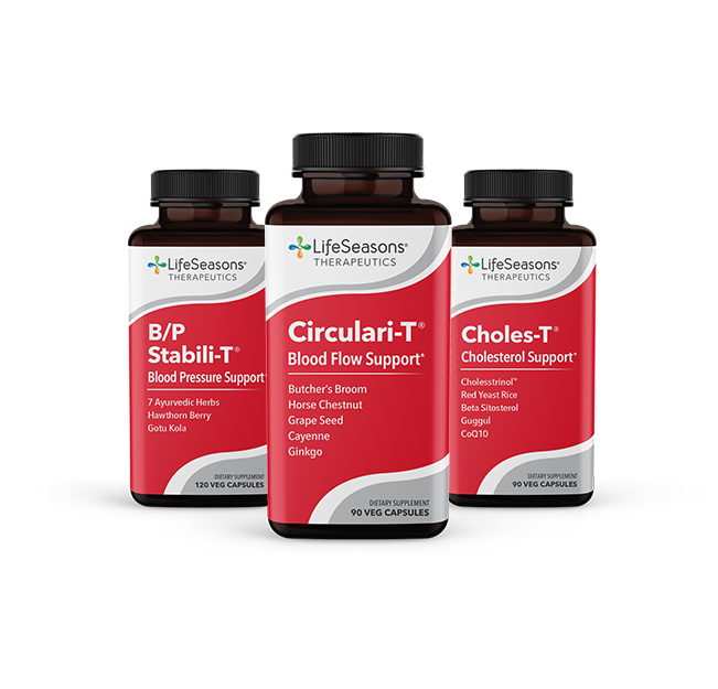
The western medical community recognizes the clinical usefulness of natural prevention in the management of macular degeneration. The current recommended supplements for dry macular degeneration include naturally occurring vitamins, pigments, antioxidants, and Omega 3 oils (8,9).
Once macular degeneration progresses to include neovascularization, the treatment drastically shifts to more aggressive options, the mainstay of which is injections into the eye. An injection of anti-vascular endothelial growth factor (Anti-VEGF) targets the protein primarily responsible for the growth of new, problematic blood vessels. Less commonly, abnormal blood vessels are targeted and destroyed using photodynamic therapy or laser surgery (9).
NATURAL WAYS TO SUPPORT EYE HEALTH
Lifestyle Practices to Support Eye Health:
- Avoid tobacco smoke (the toxins from smoke damage the retina by activating an inflammatory response) (9)
- Wear sunglasses (UV light damages the eye in the same way it can damage skin)
- Increase dietary fruits (10)
- Increase dietary nuts (10)
- Increase dietary dark green vegetables (these supports healthy retinal pigment) (10)
- Eat whole grains (10)
- Maintain a healthy weight (prevents chronic inflammatory states and hypertension) (9)
- Have regular eye examinations
Natural Supplements That Support Eye Health:
- Lutein (a natural pigment/antioxidant in the retina)
- Zeaxanthin (a natural pigment/antioxidant in the retina)
- Omega 3 fatty acids- EPA and DHA (anti-inflammatory)
- Zinc (antioxidant)
- Vitamin C (antioxidant)
- Vitamin E (antioxidant)
- Copper (11)
REFERENCES
- Isaacson W. Leonardo Da Vinci. Simon & Schuster. 2017.
- Gupta OP, Brown GC, Brown MM. Age-related macular degeneration: the costs to society and the patient. Curr Opin Ophthalmol. 2007;18:201-205.
- Risk factors for macular degeneration. American Macular Degeneration Foundation. https://www.macular.org/risk-factors. Accessed March 21, 2018.
- Coleman HR, Chan CC, Ferris FL III, Chew EY. Age-related macular degeneration. Lancet. 2008;372(9652):1835-1845. doi:10.1016/S0140-6736(08)61759-6.
- Klein R, Klein BEK, Linton KLP. Prevalence of age-related maculopathy: the beaver dam eye study. Ophthalmology. 1991;99: 933-943. doi:10.1016/S0161-6420(92)31871-8.
- Leibowitz Hm, Krueger DE, Maunder LR, et al. The framingham eye study monograph. Surv Ophthalmol. 1980;24:335-610.
- Velez-Montoya R, Oliver SC, Olson JL, Fine SL, Quiroz-Mercado H, Mandava N. Current knowledge and trends in age-related macular degeneration: genetics, epidemiology, and prevention. Retina. 2014;34(3):423-41. doi:10.1097/IAE.0000000000000036.
- Macular degeneration treatments. American Macular Degeneration Foundation. https://www.macular.org/treatments. Accessed March 22, 2018.
- Facts about age-related macular degeneration. National Eye Institute. https://nei.nih.gov/health/maculardegen/armd_facts. Accessed March 22, 2018.
- Merle BM, Silver RE, Rosner B, Seddon JM. Adherence to a mediterranean diet, genetic susceptibility, and progression to advanced macular degeneration: a prospective cohort study. Am J Clin Nutr. 2015;102(5):1196-206. doi:10.3945/ajcn.115.111047.
- AREDS2 Research Group, Chew EY, Clemons T, SanGiovanni JP, Danis R, Domalpally A, McBee W, Sperduto R, Ferris FL. The age-related eye disease study 2 (AREDS2): study design and baseline characteristics (AREDS2 report number 1). Ophthalmology. 2012;119(11):2282-9. doi:10.1016/j.ophtha.2012.05.027.